۰۳۱۳۴۷۱۷۷۰۱

چالش تشخیص بیماری های ماکولای چشم از روی تصاویر OCT

کمیته علمی :  خانم دکتر پریسا قادری دانشمند، آقای دکتر ربانی، دکتر ازهر زم، آرشام حمیدی، محمدرضا عمانی،دکتر مریم منعمیان، ریحانه رستمیان، مرضیه مختاری، دکتر سحر جرجندی، دکتر فرناز صدیقین، خانم دکتر مهنوش تاجمیرریاحی، خانم دکتر زهرا امینی 

معرفی چالش

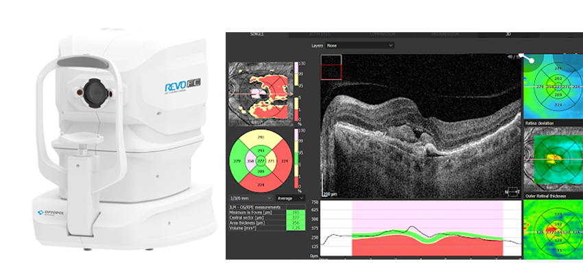
ماکولا ناحیه ی مرکزی کوچک از شبکیه چشم است که تیز بینی (VA) را کنترل می کند. توانایی خواندن ، تشخیص صورت ها ، رانندگی ، تماشای تلویزیون ،استفاده از کامپیوتر و انجام دادن هر کار بینایی که نیازمند دیدن جزییات باشد با سلامت ماکولا تعیین می شود. هدف این چالش تشخیص بیماری های ماکولا از روی تصاویر OCT چشم انسان است. بیماری های ماکولا مورد نظر شامل ورم ماکولای دیابتی(DME)، تخریب ماکولای وابسته به سن(AMD)، نئوواسکولاریزیشن کروئیدال (CNV) و سوراخ ماکولا (MH) می­باشد. در تصویربرداری های صورت گرفته، طول موج مرکزی، پهنای باند طیفی و نرخ  A-SCAN مربوط بهSS –  OCT به ترتیب 1064 نانومتر، 100 نانومتر و 100 کیلوهرتز بوده است.

در داده های این چالش افراد مبتلا به DME با برچسب “1” (73 نفر)، افراد سالم با برچسب “0” (54 نفر)، و بیماران غیر دیابتی با اختلالات AMD، CNV، یا سوراخ ماکولا (MH) با برچسب “2” (64 نفر) نمایش داده شده اند. هر فرد شامل 300 B-scan  با وضوح 300 تا 1200 در 300 پیکسل است. این تصاویر در مجموعه داده با عنوان “1- Raw Dataset ” موجود است.

از آنجایی که برخی از داده ها کیفیت پایینی دارند یا ناحیه ماکولا ضبط نشده است، ارزیابی کیفیت (QA) بر روی تصاویر مرحله قبل انجام شده است تا تعیین کند که آیا B-scan برای تجزیه و تحلیل های بیشتر مناسب است یا خیر. برای مرحله آماده سازی داده ها، ارزیابی کیفی دستی انجام شد. داده ها به دو دسته تقسیم شدند: 190 پوشه از B-scan های واجد شرایط و 188 پوشه از B-scan های غیر واجد شرایط. هر پوشه شامل 1 تا 300 B-scan است. و همه B-scan ها در هر پوشه به یک فرد خاص تعلق دارند. داده های آموزشی برای QA در ماتریس “0-QA-data” موجود است. برای آموزش QA از روش Deep Convolutional Neural Network  استفاده شده است. در مرحله بعد از QA برای ارزیابی کیفیت تصاویر مجموعه ” 1-Raw Dataset ” استفاده شد. و افراد با بیش از 30  B-scan واجد شرایط انتخاب شده اند.

ساختار داده ها:

1-Raw Dataset: شامل 191 پوشه از تصاویر خام است. هر پوشه مربوط به یک فرد شامل 300 B-scan  با وضوح 300 تا 1200 در 300 پیکسل است.

2-Raw Dataset QA: شامل 124 پوشه است که پس از ارزیابی کیفیت تصویر با برش خودکار به دست آمده است. هر سوژه شامل 30 تا 300 B-scan  با وضوح 300 در 300 پیکسل است.

3-Enhanced Dataset QA: شامل 124 پوشه است که پس از افزایش کنتراست تصویر با استفاده از تبدیل گاوسی و نرمال سازی روی ”2- Raw Dataset QA” به دست آمده است.

4-Denoised dataset QA: شامل 124 پوشه است که پس از حذف نویز و نرمال سازی بر روی تصاویر “3-Enhanced Dataset QA”  به دست امده است.

:Aligned Dataset QA شامل 124 پوشه است که پس از تراز کردن تصویر با کمک لایه ILM با استفاده از روش نیمه اتوماتیک و نرمال سازی بر روی  “4-Denoised dataset QA”به دست آمده است.

OCT Basel Data

The central wavelength, spectral bandwidth and A-scan rate of the custom-made SS-OCT are 1064 nm, 100 nm, and 100 kHz, respectively.

The participants included people with DME or “1” (73 subjects), Healthy or “0” (54 subjects), and Non-diabetic patients or “2” (64 subjects) with AMD, CNV, or Macular Hole (MH) disorders. Each subject contains 300 B-scans with 300 to 1200 x 300 pixels resolution. These 191 are available in “1-Raw Dataset”.

Since some of the data have poor quality or the macular region was not captured, Quality Assessor (QA) was designed to automatically determine if the B-scan is suitable for further analyses or not. For the data preparation step, the manual quality assessment was performed on 191 volumes. The data was divided into two categories: 190 folders of qualified B-scans and 188 folders of non-qualified B-scans. Each folder consists of 1 to 300 B-scans. And all B-scans in each folder belong to one specific subject. The training data for QA is available in “0-QA-data”. Deep Convolutional Neural Network 1 method is employed to train QA. In the next step, QA was used to assess the quality of subjects in “1-Raw Dataset”. Volumes with more than 30 qualified B-scans passed.

0-QA-data: This data includes 190 folders of qualified B-scans and 188 folders of non-qualified B-scans. Each folder consists of 1 to 300 B-scans with 300 x 300 pixels resolution.

1-Raw Dataset: This dataset includes all 191 volumes before any processing. Each volume contains 300 B-scans with 300 to 1200 x 300 pixels resolution. The participants included people with DME or “1” (73 subjects), Healthy or “0” (54 subjects), and Non-diabetic patients or “2” (64 subjects) with AMD, CNV, or Macular Hole (MH) disorders.

2-Raw Dataset QA: Includes 124 volumes which has been obtained after image quality assessment with automatic cropping. Each subject contains 30 to 300 B-scan with 300 x 300 pixels resolution. The participants included people with DME or “1” (40 subjects), Healthy or “0” (50 subjects), and Non-diabetic patients or “2” (34 subjects) with AMD, CNV, or Macular Hole (MH) disorders.

3-Enhanced Dataset QA: Includes 124 volumes which has been obtained after image contrast enhancement using Gaussianization transformation, and normalization on “2-Raw Dataset QA”. The participants included people with DME or “1” (40 subjects), Healthy or “0” (50 subjects), and Non-diabetic patients or “2” (34subjects) with AMD, CNV, or Macular Hole (MH) disorders.

4-Denoised dataset QA: Includes 124 volumes which has been obtained after image denoising2, and normalization on “3-Enhanced Dataset QA”. The participants included people with DME or “1” (40 subjects), Healthy or “0” (50 subjects), and Non-diabetic patients or “2” (34 subjects) with AMD, CNV, or Macular Hole (MH) disorders.

5-Aligned Dataset QA: Includes 124 volumes which has been achieved after image alignment based on Internal Limiting Membrane (ILM) using the semi-automatic method, and normalization on “4-Denoised Dataset QA”. The participants included people with DME or “1” (40 subjects), Healthy or “0” (50 subjects), and Non-diabetic patients or “2” (34 subjects) with AMD, CNV, or Macular Hole (MH) disorders.